X-RAY CHECKS ON PAINTINGS: REVOLUTIONIZING ARTISTIC VISION English Version

Pubblicato da Brigida Michele in Xrayconsult · Lunedì 19 Feb 2024
Tags: Raggi, X, Dipinti, Ripensamenti, Visione, ai, raggi, X, Controlli, raggi, X, Segreti, artistici, celati
Tags: Raggi, X, Dipinti, Ripensamenti, Visione, ai, raggi, X, Controlli, raggi, X, Segreti, artistici, celati
X-RAY CHECKS ON PAINTINGS:
REVOLUTIONIZING ARTISTIC VISION
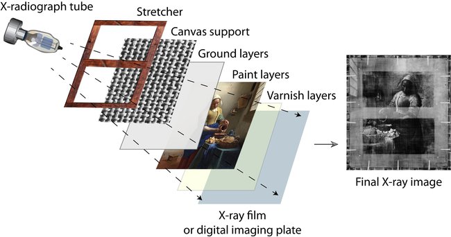
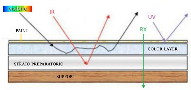
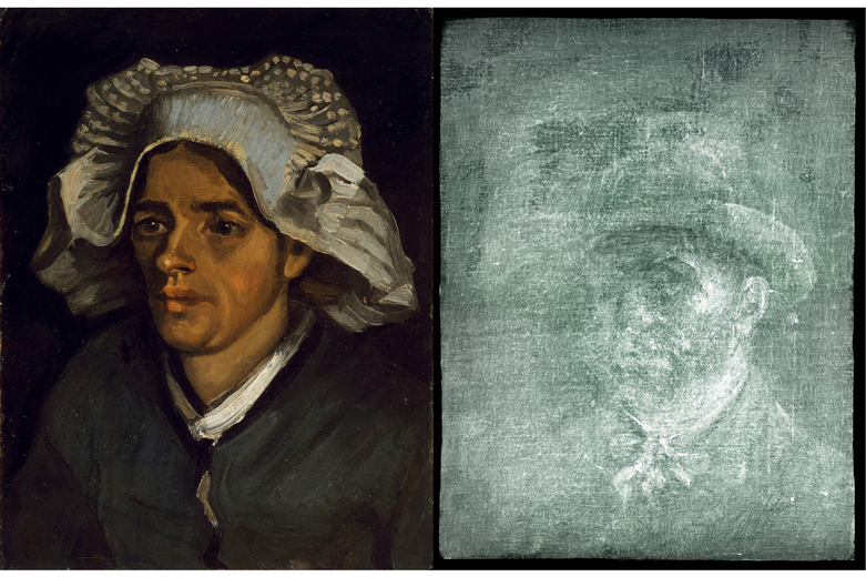
Chapter 1: Introduction to X-ray Inspections on PaintingsPaintings serve as extraordinary testimonies to human creative genius, providing a window into the past and the minds of artists.However, a painting's surface often conceals hidden secrets and details that can reveal much more than what is visible in history.Layers of paint and pigments hide reconsiderations, changes, and decisions that enrich the understanding of the artistic process.X-ray inspections emerge as a revolutionary technique to explore the concealed secrets beneath the surface of paintings.This non-invasive methodology allows penetrating the deeper layers of the artwork, unveiling details invisible to the human eye.The interaction of X-rays with various materials in the painting generates a radiographic image that highlights its internal structure, offering a complete and detailed view of the artwork.The importance of exploring the deep layers of paintings through X-ray inspections lies in revealing hidden artistic secrets.These technological tools enable the analysis of the artist's reconsiderations, changes in intent, and stylistic choices by visualizing the different layers of work.Moreover, they facilitate evaluating the conservation status of the artwork and planning targeted and conservative restoration interventions.Original Painting Painting X-Ray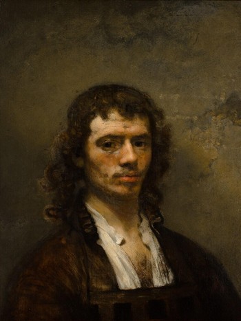
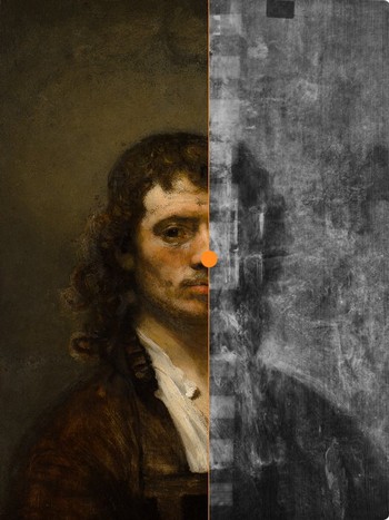 The X-ray of the renowned Self-Portrait by Carel Fabritius (1205) reveals how the artist initiallyportrayed himself with a white collar before choosing the open shirt we see now.Currently, the painting is housed at the National Gallery of Art in Washington, D.C.1.1 Color Density and Radiography: An In-depth Analysis of the Chemical-Physical Properties of PigmentsIntroduction:Radiographic examination of paintings represents an invaluable non-invasive analysis method, capable of revealing details otherwise inaccessible to the human eye.Among the peculiarities of this technique stands out its ability to discern the various pigments used by the artist based on their heterogeneity in terms of radiographic density.This variability, in turn, has a direct correlation with the chemical-physical composition of the pigments themselves.1.2 Chemical-Physical Composition and Radiographic Density:Pigments, the colored powders used for paint production, were historically derived from natural raw materials such as minerals, earth, and plants.Their chemical-physical composition varied significantly based on the nature of the starting material, determining a different radiographic density.

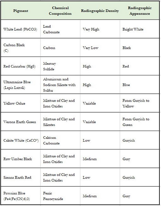
Examples of Natural Pigments and Radiographic Density:
- White: Lead white (PbCO3), characterized by high density, appears radiopaque (white) in radiographs. Lime white (CaCO3), with lower density, takes on a grayish tone.
- Black: Carbon black (C), being a lightweight pigment, is radiotransparent (black) in radiographs. Raw umber, composed of a mixture of clay and iron oxides, has a higher density and appears grayer.
- Red: Cinnabar red (HgS), with high density, appears radiopaque in radiographs. Sienna red, composed of clay and iron oxides, with lower density, takes on a grayish tone.
- Blue: Ultramarine blue (lapis lazuli), characterized by high density, appears radiopaque in radiographs. Prussian blue (Fe4[Fe(CN)6]3), with lower density, appears grayer.
1.3 Artisanal Processes and Impurities:In the past, pigment production was a complex artisanal process imbued with variables.Impurities in natural raw materials and fluctuations in production processes influenced the chemical-physical composition of pigments and, consequently, their radiographic density.
Example:Lead white, obtained by grinding galena mineral (PbS), could contain traces of other minerals like barite (BaSO4).Barite, being a pigment of high density, could generate more intense radiopaque areas in radiographs.The combined use of radiography and chemical-physical analysis of pigments represents an invaluable method for understanding, conserving, and restoring paintings.
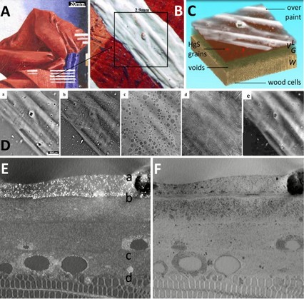
X-ray Images in X-ray Computed Laminography TechniqueA: Visual photograph; B: close-up of A, showing a brushstroke of white lead covering the red surface of the painting; C: 3D rendering of the data, displaying the reconstructed volume; D: series of virtual laminographic sections at, parallel to the surface of wood/paint at the depth indicated in E: (a) surface white lead, (b) particles of pigment in red paint; (c) spherical voids in the basal layer; (d) cells in the wooden support; panel (e) shows the corresponding X-ray of the volume displayed in C; E, F: virtual cross-sectional views obtained by applying a maximum (E) or minimum (F) filter of 750 images, oriented perpendicularly to the axis of wood cell structures.
Other factors that can influence radiographic density:
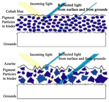
- Particle Size of Pigments: Pigments with a finer particle size tend to be more radiopaque.
- Color Application Technique: A color applied more thickly appears more radiopaque than one applied more thinly.
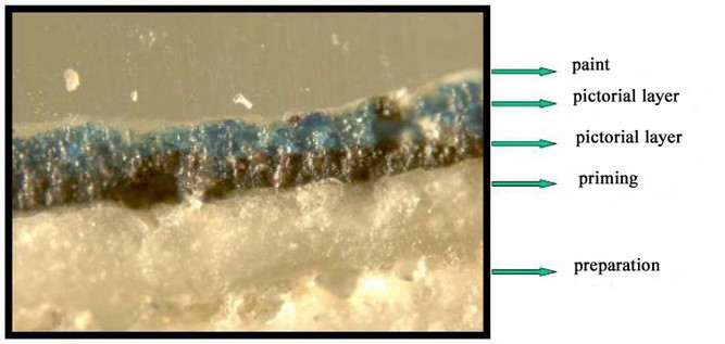
- Preparation Layer: The preparation layer, also known as priming or gesso, represents a fundamental component of a painting.
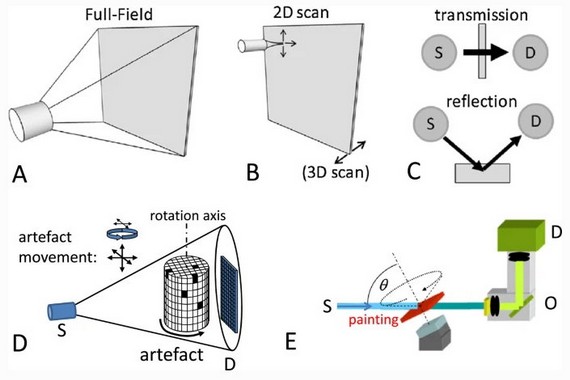
2.1 Overview of X-ray Technologies:
There are various types of X-ray systems, each with specific characteristics and advantages:2.1.1 Traditional Radiography:- Utilizes an X-ray source and a radiographic plate to capture the image.
- Provides high resolution and is effective for detecting structural details and pigment composition.
- Requires development time and involves prolonged radiation exposure, imposing limits on flexibility and potential damage to the artwork.
2.1.2. Digital Radiography:
2.1.4. Optical Coherence Tomography (OCT):
The choice of the most suitable radiography system depends on various factors, including:
- Type of artwork: Materials, dimensions, and conservation status.
- Desired information: Internal structure, composition of pigments, pentimenti, underlying paintings.
- Available resources: Budget, time, and specific skills.
The use of X-ray systems represents a continuously evolving research field contributing to expanding knowledge and possibilities for the study and conservation of artistic heritage.Introduction:Radiographic investigation proves to be an invaluable methodology for the non-invasive analysis of paintings, providing valuable information on their internal structure, chemical composition, state of preservation, and painting technique without causing damage.2.3 Fundamental Technical Parameters:2.3.1. Penetrability:The ability of X-rays to penetrate a paint layer depends on their energy, measured in keV (kilo-electron volts). Paintings with thickness or high density, such as those on panels or with a rich paint impasto, require higher energy (kV) for adequate penetration and the acquisition of meaningful diagnostic information.
Example:The radiographic analysis of a 16th-century panel painting with a thick gesso ground would require an X-ray system with an energy of at least 150 keV to obtain a detailed radiograph of the internal structure and any underlying preparations or pentimenti.
2.3.2. Spatial Resolution:The ability to distinguish fine details, such as canvas texture, brushstrokes, and cracks, depends on the size of the X-ray source focal spot and the digital detector.A smaller focal spot and a high-resolution detector provide better image sharpness, allowing the identification of micro-structures and minimal details.
Example:The radiographic analysis of a miniature painting on parchment would require an X-ray system with a focal spot size below 50 µm and a high-resolution detector to highlight details such as fine lines, highlights, and incisions.
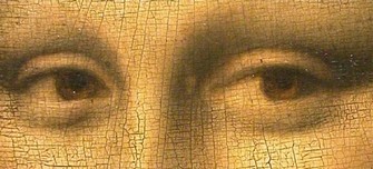
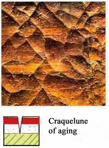
"Craquelure on the Mona Lisa"Craquelure, or crettature, represents a delicate web of fractures that emerges on diverse materials' surfaces.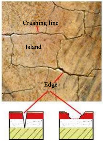
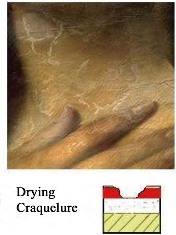 Craquelure in Paintings and Contrast Dynamics in X-ray Radiography Craquelure is a common phenomenon in paintings, especially oil ones.It occurs when the paint layer dries and contracts, leading to the formation of a series of superficial cracks.The type of craquelure can vary based on the painting technique, materials used, and the time elapsed since the creation of the artwork.2.3.3. Contrast Dynamics:The ability to distinguish between different pigments, binders, and materials, such as canvas, chalk, and wood, depends on the grayscale range of the radiographic image.A system with a wide range of grays allows for highlighting details and nuances that might be otherwise invisible. This facilitates stratigraphic analysis and the differentiation of gaps, defects, and restorations.
Example:The radiographic analysis of a 19th-century oil painting on canvas would require an X-ray system with a broad grayscale range to differentiate various color tones, glazes, and any modifications made by the artist during the creation process.
"Painting with Various Glazes""Velatura" is a painting technique involving the application of a layer of fresh (or wet) transparent or semi-transparent color over a previously dried layer. Its purpose is to enhance the brightness and transparency of a painting's tones.2.3.4. Radiation Dose:Exposure to radiation must be minimized to avoid damage to the artwork and protect the health of operators.X-ray systems with low radiation emission and rapid acquisition times are preferable, ensuring the safety and efficiency of the analysis process.

Example:The use of an X-ray system with digital technology and dose optimization software could reduce radiation exposure by up to 50% compared to traditional systems, ensuring the safety of both the painting and operators.
2.4 Interaction with the Painting:X-rays interact with the painting in various ways:2.4.1. Absorption:Denser materials, such as lead present in white pigments, absorb more X-rays, creating darker areas in the radiographic image.This provides information about density distribution, the presence of inclusions, and the painting technique used.
Example:In the radiographic image of a 17th-century painting, darker areas would correspond to regions with a higher concentration of lead white pigment (biacca), used to create highlights and areas of light.
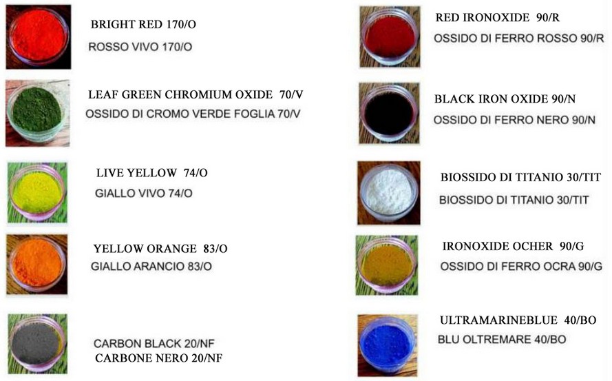
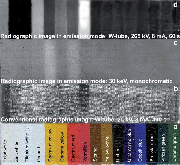
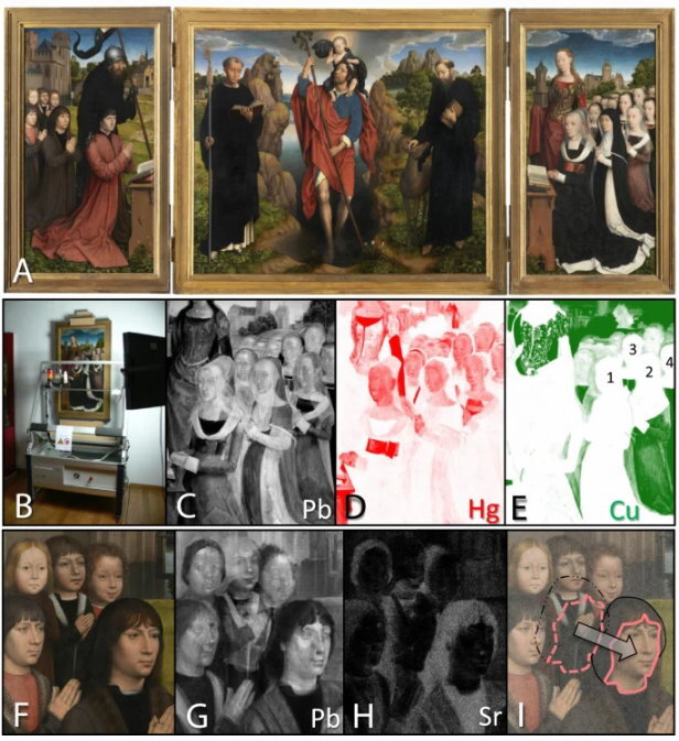
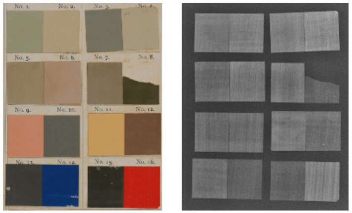
Artists traditionally used lead-based paints for a wide range of colors, including whites.In the illustration, the first image (top) depicts the visual appearance of various lead-containing paints on paper under normal light.The second image (bottom), obtained through radiography, not only reveals brushstroke traces but also highlights the presence of heavy elements like lead.For instance, the red point in the bottom right corner (no. 16) appears brighter in the radiograph compared to the dark/ blackish point (no. 15) on its left.This difference suggests a higher concentration of lead in the red paint.2.4.2. Diffusion:X-rays can be scattered by particles within the painting material, creating a "fog" effect in the image. Diffusion is useful for analyzing microstructures, such as canvas texture or pigment granularity.
Example:Radiographic analysis of a 20th-century painting on jute canvas could highlight the canvas texture through X-ray diffusion, providing information about the weave and preparation of the support.
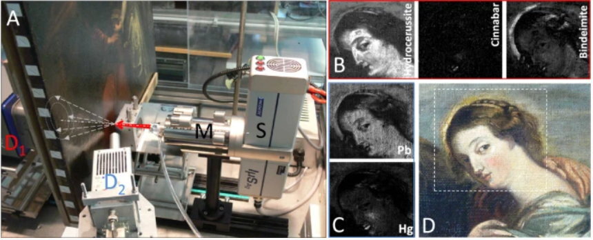
2.4.3. X-ray Fluorescence (XRF):XRF analysis in the characterization of artistic pigments. Some components in the artwork, like copper in malachite green pigment or iron in Sienna earth pigments, generate distinctive fluorescent radiation when exposed to X-rays.This fluorescence offers detailed mapping of chemical elements in the artwork, avoiding the need for invasive material sampling.XRF provides real-time information on the entire paint layer thickness. Comparing results visually with the present colors allows associating many elements with different pigments.For instance, the presence of mercury in a red hue indicates the use of cinnabar. This unique property of XRF enables accurate identification of pigments, binders, and other materials, significantly contributing to understanding the artistic technique and history of the artwork.
Example:Radiographic analysis of a 14th-century mural might use fluorescence to identify the presence of lapis lazuli-based pigments, a precious and rare material used for achieving intense blue color.
 2.5 Relevant Considerations:
- The choice of the most suitable X-ray system depends on the specific characteristics of the painting, the desired information, and the objectives of the analysis.
- It is crucial to use the minimum radiation dose necessary to obtain a diagnostically quality image, balancing the need for information with the protection of the artifact.
- Highly qualified and experienced personnel are necessary for the use of X-ray systems and the interpretation of images, ensuring the proper execution of the analysis and the reliability of results.
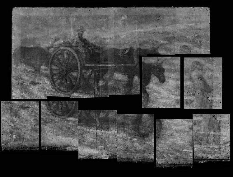
Chapter 3: Artist's Significant Discoveries through RadiographyRadiography is an image generated by X-rays, capable of penetrating bodies based on density, thickness, and atomic weight.This technique reveals the internal structure of paintings, providing details about the composition of the support (type and weave of the original canvas, type of wood and assembly of boards, etc.) and the characteristics of preparatory layers, sketches, and paint layers.In artworks, lead white pigment (biacca), common in sketches, highlights, and chromatic mixtures, appears more opaque to X-rays (radiopaque).Conversely, light strokes, protective layers, and varnishes do not provide detectable information.Radiographic analysis highlights crucial aspects for conservation and restoration, such as canvas damage, wood support infestations, and injuries, loss of paint layers or surface film, characteristics of integrations, and relationships with deficiencies.3.1 Underlying Paintings:
Madonna del Rosario by Caravaggio: The radiographic examination has revealed significant details, such as the painting technique similar to other Caravaggesque works and the presence of a preceding support canvas, aligning with the proposed dating.The initial X-rays of the painting date back to 1950, following a restoration due to theft that had damaged the canvas. The radiographs identified the presence of an underlying composition, featuring a figure of a saint not present in the final version. This discovery led to new hypotheses about the genesis of the painting and Caravaggio's creative process.In 2008, during a subsequent restoration, the painting underwent a more in-depth radiographic analysis using advanced digital technologies. The new radiographs provided high-resolution images that revealed additional details of the underlying composition, including the presence of a landscape and some angelic figures.
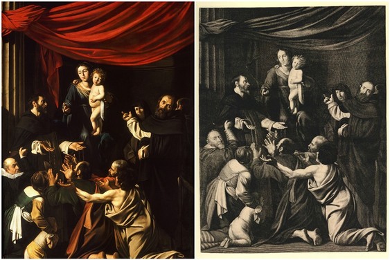
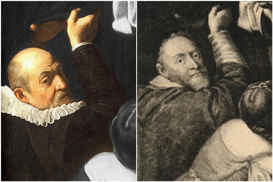
Original Painting X-ray of the painting Detail of the Painting X-ray Reveals a Change in SubjectPainting by Caravaggio - "The Madonna of the Rosary" dating back to 1607"La Miséreuse Accroupie" by Picasso, painted in 1902 during his "blue period," depicts a crouched woman wrapped in a cloak.This masterpiece is renowned for Picasso's distinctive use of intense blue, characteristic of his artistic style during that period.The portrayed woman conveys sadness and poverty, reflected in the chromatic choices and expression of the subject.The painting's surface conceals another underlying artwork, revealed through radiographic analysis in 1992, adding mystery and complexity to the piece.
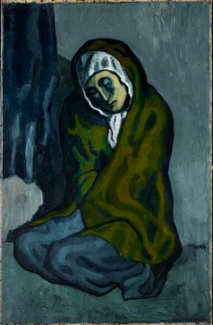
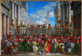
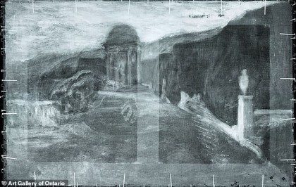 Original Painting Spectrometry of the Painting Analysis of the paintingPicasso's "La Misereuse Accroupie" (The Crouching Woman)was created during his Blue Period, characterized by the predominant use of blue and green hues.Radiographic examinations of Picasso's painting revealed hidden artworks beneath the visible surface.Beneath the canvas of the 1902 painting from Picasso's Blue Period, another artwork by the same artist and a landscape are suspected to exist.This discovery provides an intriguing perspective on Picasso's artistic practice and stylistic evolution over the years.3.2 Overlapping Paintings:Paolo Veronese's "The Wedding at Cana" underwent radiographic analysis, uncovering two compositions beneath the final work.This offers insight into Veronese's creative process, showcasing his skill in modifying and reworking his pieces.The initial X-rays date back to 1930, around fifty years after the painting's restoration in 1882-1883.Subsequent radiographs in the '50s and '60s, during additional restoration, deepened understanding of Veronese's painting technique, revealing figures underlying the final composition.In 2008, during a significant exhibition dedicated to the Venetian painter, the artwork underwent a comprehensive radiographic investigation using advanced digital technologies.The new high-resolution radiographs provided unprecedented detail, uncovering previously unknown details and pentimenti.

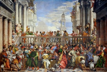
 Original Painting X-ray of the painting Cutting of the Canvas by French Soldiers"The Wedding at Cana" by Paolo Veronese is a canvas created to adorn the wall of the monastery's refectoryin the Basilica of San Giorgio Maggiore in Venice. The artwork is currently housed in the Louvre.
"The Old Guitarist" by Pablo Picasso: The painting in question was created during his Blue Period, between late 1903 and early 1904.Initially depicting an elderly guitarist, X-ray analyses revealed an underlying female figure.It is believed that Picasso started by painting a portrait of a woman, later covering it with the guitarist's figure. X-ray images clearly show the outlines of the female figure beneath the upper layer of the painting.This analysis led to the discovery of the previously hidden female presence beneath the painting's surface.X-ray exploration served as a non-destructive technique to deepen the understanding of Picasso's artistic evolution and reveal intricate details.
Original Painting X-ray of the painting Restored Hidden Image"The Old Guitarist" by Pablo Picasso;Currently, the painting is housed at the Art Institute of Chicago in the United States.

3.3 Restoration Interventions:Although initially little known, the use of X-rays in art emerged shortly after their discovery, originating in the medical field.From there, the technology expanded to anthropology and mummy studies, eventually reaching the analysis of paintings.One of the first paintings subjected to such analysis was "The Deposition from the Cross" by Van Der Weyden in the 1970s.Over time, the use of X-rays to reveal the artistic past has become commonplace in institutes, museums, and galleries worldwide.
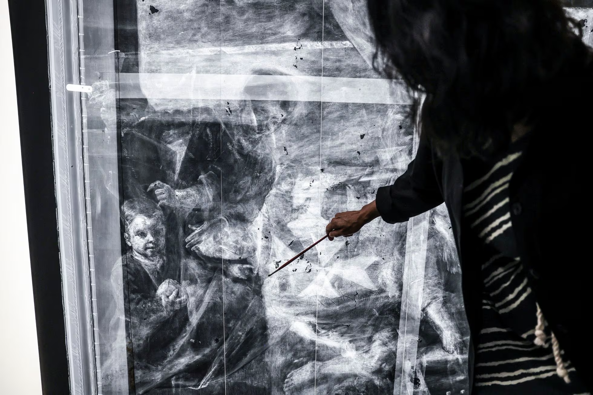 Restorer at workX-rays unveil details of every layer in works of art, such as paintings and sculptures, providing information about primers, support, and materials used.However, interpretation requires expertise due to data overlap, potentially leading to misconceptions. Nevertheless, X-rays remain fundamental tools for revealing hidden details.To unveil the secrets of artworks, it's essential to transport large paintings to the laboratory. Here, specialists carefully position them, calculate the required film quantity, and apply it with precision to avoid detail losses.This approach demands accuracy and attention to accurately reveal the hidden elements in artworks.Chapter 4: Artist's Rethinking: A Journey into Creative PastExploring the creative process offers a fascinating opportunity to deepen understanding of the artist and their aesthetic choices.Paintings, with their enigmatic surface, conceal secrets and stories waiting to be uncovered.Thanks to the use of radiographic technologies, the possibility arises to cross the threshold of the visible and embark on a journey through time to discover the artist's rethinkings.4.1 Assuming the role of a window into the past, radiography allows:
- Analyzing changes made during the painting process: pentimenti, reconsiderations, and changes the artist made on the canvas.
- Understanding the evolution of the creative process: how the initial idea developed and transformed into the final version.
- Identifying employed painting techniques: layer overlaps, glazes, incisions, and other details otherwise invisible to the human eye.
In this chapter, we will venture into an analysis of emblematic cases illustrating how the use of radiographic technologies allows exploring the artist's rethinkings and gaining a deeper knowledge of their working method.4.2 Example of pentimenti and corrections:"The Tempest" by Giorgione: Radiographic analysis of the painting "The Tempest" has revealed several changes made by the artist during the creation phase. In particular, the following pentimenti have been identified.Original Painting X-ray of the painting
"La Tempesta di Giorgione"The painting is currently housed in the Gallerie dell'Accademia in Venice, Italy.
Giorgione's alterations to "La Tempesta" provide a valuable opportunity to observe his creative process in action. The pentimenti and corrections highlight his attention to detail, constant pursuit of balance and harmony, and his skill in modifying and improving his works.In addition to this example, several other artworks have undergone radiographic analysis, revealing significant pentimenti and corrections.Among these, we remember:"Il Cenacolo" of Leonardo da Vinci: The radiographic examination identified the presence of an apostle initially depicted in place of Judas Thaddeus."Il Cenacolo di Leonardo da Vinci"Currently, the painting is housed in the refectory of the Convent of Santa Maria delle Grazie in Milan, Italy.Originally, it was depicted seated in the place of Judas Thaddeus, but later, Leonardo painted over it.The reason for this change is not known, but it is believed that Leonardo may have decided to alter the composition of the fresco for theological or artistic reasons.The radiographic examination that revealed the presence of the original figure was conducted in 1978.X-rays showed that the figure of Saint Peter had been painted using a technique different from that used for the other figures in the fresco.This suggests that the figure of Saint Peter was added later, after the fresco had already been completed.

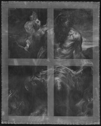
"San Girolamo" by Antony Van Dyck: Anthony van Dyck (1615) initially used the canvas on which he painted his magnificent Saint Jerome for a painting of a reclining nude nymph.He rotated the canvas from landscape to portrait format before starting his representation of the saint. The arm of the nymph is clearly visible on X-rays.
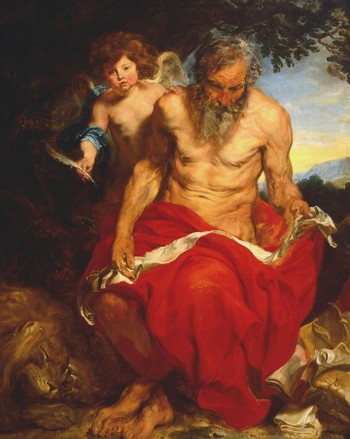
 Original Painting X-ray of the painting"San Girolamo di Antony Van Dyck"Currently, the painting is housed at the Boijmans Van Beuningen Museum in Rotterdam,.Il dipinto di Rembrandt intitolato "L'attesa di Tobia e Anna" è un'opera autografa realizzata con tecnica ad olio su tavola nel (1659).
Le dimensioni del dipinto sono di 40,5 x 54 cm. Quest'opera rappresenta una scena tratta dalla storia biblica di Tobia e Anna.Il dipinto di Rembrandt intitolato "L'attesa di Tobia e Anna" è un'opera del 1659 che raffigura l'anziano Tobia e sua moglie Anna che attendono il ritorno del loro figlio Tobiolo dalla Media.Il dipinto è olio su tavola ed è attualmente conservato al Museo Boijmans Van Beuningen di Rotterdam.

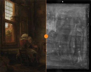 Original Painting X-ray of the painting"The Waiting of Tobias and Anna"by Rembrandt Currently, the painting is housed at the Boijmans Van Beuningen Museum in Rotterdam, Netherlands.It appears that Rembrandt painted Tobias and Anna against a still life backdrop.The underlying motives are invisible to the naked eye and were only uncovered through recent radiographic techniques.Radiographs revealed that beneath the main scene of Tobias and Anna, there is a still life composition featuring various objects, including:
- A table with a tablecloth
- A basket of fruit
- A glass of wine
- A loaf of bread
- A knife
The reason why Rembrandt painted a still life beneath the main scene is not clear. Some hypotheses include:
- Rembrandt was experimenting with different compositions.
- Rembrandt used the still life as filler for the canvas.
- The still life carries symbolic meaning.
The still life might symbolize the everyday life that Tobias and Anna left behind when Tobias departed for Media.It could also be a symbol of divine providence, watching over them in their anxiety and concern.The discovery of the still life under "The Waiting of Tobias and Anna" is a significant contribution to our understanding of Rembrandt's creative process.It provides a new perspective on this iconic painting and helps us better comprehend his working methods.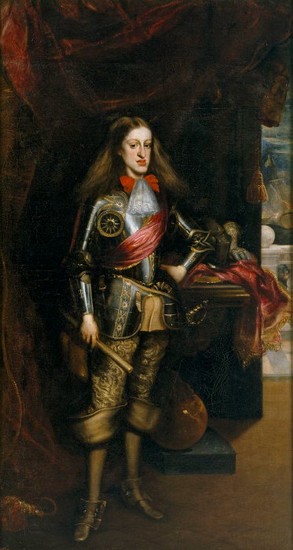
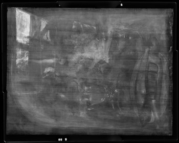
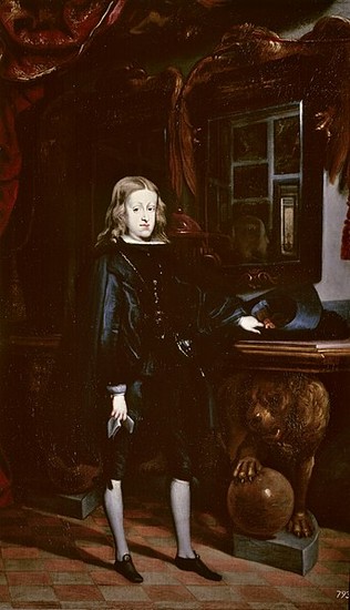
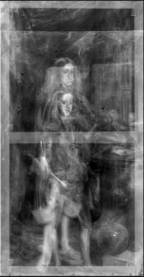 Original Painting X-ray of the painting Another painting from the Museum of Asturias,Carlo II at the age of ten."L'Incantato di Juan Carreño de Miranda"Currently, the painting is housed in the Prado Museum in Spain.This portrait of the adult Carlos II, painted by Carreño de Miranda in 1681, conceals another artwork.Carreño reused a canvas on which he had previously painted a portrait of the king when he was younger.The Evolutionary Portrait of Carlos II "The Enchanted" by Juan Carreño de Miranda, currently housed at the Museo del Prado.Originally, the painting bore similarities to another work stored at the Museum of Fine Arts of Asturias, revealed through an X-ray exhibited by the Museo del Prado on the anniversary of Marie Curie's death.Contrary to speculations, the preserved Asturian painting represents Carreño de Miranda's initial portrait of the young monarch titled "Carlos II at the age of ten." The young king is depicted with long blond hair, dressed in Spanish simplicity, adorned with the Golden Fleece, leaning on a table with a hat in hand and clutching a letter.A later version at the Museo del Prado, titled "Carlos II in Armor" from 1681, portrays the king in adulthood, wearing armor, maintaining the same posture but in a different context.The X-ray reveals the presence of another portrait similar to the first, hidden beneath the later work, depicting Carlos II at the age of ten.Chapter 5: Implications for Art HistoryThe introduction of radiographic inspections has revolutionized the way we study and understand works of art.This non-invasive technology allows us to look beyond the surface of a painting, unveiling secrets and hidden stories that have remained concealed for centuries.In this chapter, we will examine the implications of using X-rays for art history, particularly in three areas:5.1. Restoration:
- Conservation Analysis: X-rays can identify damages, cracks, deformations, and other structural issues not visible to the naked eye.
- Restoration Guidance: Radiography provides valuable information about materials used, painting techniques, and any modifications made to the artwork over time.
- Efficacy Monitoring: X-rays can be employed to monitor the progression of damages and the effectiveness of restoration efforts.
5.2. Attribution and Dating:
- Comparison with Reference Works: Radiographic analysis aids in attributing a painting to a specific artist or historical period by comparing its characteristics with reference works.
- Dating of the Painting: X-rays can be used to date the wooden supports and other materials used in the creation of the painting.
Radiography is a fundamental tool in dating paintings, especially those on wooden supports.This technique delves into the deeper structure of paintings, revealing details about the composition of materials used.For instance, in the conservation of painted wooden supports, the relationship between the painted panel and the artwork itself can be analyzed through radiography.Widely accepted in the fields of art and conservation, radiography helps establish the dating of artworks and understand the processes involved.Art historians, archaeologists, and restorers often use radiography as a consolidated tool to obtain detailed information about the materials employed in creating paintings.5.3. Historical-Artistic Research:
- Unveiling the Genesis of the Work: X-rays can reveal preparatory drawings, pentimenti, and other traces of the artist's creative process.
- Search for Hidden Details: Radiography can bring to light details invisible to the naked eye, such as signatures, inscriptions, or symbols concealed beneath the painting's surface.
- New Interpretations: Information obtained from radiographic analysis can contribute to formulating new interpretations and reevaluating the historical and artistic significance of a work.
The Mona Lisa, one of Leonardo da Vinci's most celebrated works housed at the Louvre, may appear as a singular entity superficially, but the art world is rich with dozens of copies and variations of this iconic masterpiece. While none of these can truly approach the genius of the master, reinterpretations abound in various forms and global contexts.The use of X-ray examinations in the artistic field has revolutionized the understanding and preservation of artworks. This technology enables the exploration of the internal structure of paintings on canvas, wood, paper, and cardboard, revealing hidden details about the materials used and the phases of creation. Through X-ray radiography, it is possible to analyze values of light and shadow, identifying restorations and contributing to an improved dating of artworks.The ability to penetrate the superficial layers of paintings unveils artistic secrets, such as preparatory drawings executed in leadpoint, and facilitates the detection of potential damage or alterations. Additionally, X-ray examinations are essential for conservation, allowing targeted interventions by removing old retouches and non-original restorations, as demonstrated in recent restoration projects.5.4 Beyond these three main areas, the use of X-rays also has other implications for art history, including:
- Advanced Imaging Techniques: Evolution in imaging techniques, such as X-ray computerized tomography (micro-CT) and X-ray fluorescence spectroscopy (XRF), promises increasingly precise information about artworks.
- Artificial Intelligence and Machine Learning: Integrating artificial intelligence and machine learning into radiographic analysis will automate the identification of invisible details, opening new research scenarios.
- Sensor Miniaturization: The miniaturization of sensors will enable non-invasive analysis directly on paintings without the need for sampling.
6.2 Applications and Future Perspectives:
- Research on Underlying Paintings: Advanced technologies will be employed to identify hidden paintings, providing new opportunities for discovery and reassessment of considered lost artworks.
- In-Depth Study of Painting Techniques: Non-invasive analysis will allow in-depth knowledge of artists' painting techniques, offering insights into materials, tools, and working methods.
- Dating and Attribution: New technologies will enhance accuracy in dating and attributing artworks.
- Unmasking Forgeries: Non-invasive analysis will aid in identifying forgeries and combating the illegal art market.
6.3 Challenges and Ethical Considerations:
- Democratic Access to Technologies: Ensuring accessibility of technologies to a wide range of scholars is essential.
- Specialized Training: Specialized training programs will be crucial for correctly interpreting non-invasive analysis data.
- Ethics and Conservation: It is fundamental that the use of technologies does not harm artworks, adopting rigorous protocols and best practices.
In conclusion, the future of non-invasive analysis holds rich possibilities, with the development of sophisticated technologies and interdisciplinary approaches that will open new horizons in the understanding and valorization of artistic heritage.Continued commitment to perfecting and responsibly applying these techniques will ensure the preservation and enjoyment of artworks for future generations.
Bibliography:1. Scoprendo l'arte con i controlli a raggi x! - Video tutorial sull'utilizzo dei raggi X nell'arte
- Autore: Maria Anna Cerini
- Anno: 2023
2. Dalla conservazione alla storia dell'arte - Approfondimento sulla diagnostica non invasiva per restauratori e conservatori
- Autore: Luigi Campanella e Francesca Castelli
- Anno: 2020
3. Radiografia d'Arte: Tecniche Avanzate di Analisi
- Autore: Piero Cazzola e Giovanni Battista Galizzi
- Anno: 2015
4. Il Futuro della Conservazione: Raggi X e Nuove Prospettive
- Autore: Irene F.M. van der Vlies e Koen Janssens
- Anno: 2024
5. Raggi X nell'Arte: Dal Passato al Futuro
- Autore: Carlo Ginzburg e Andrea Zezza
- Anno: 2018
6. L'Arte dello Svelamento: Raggi X nei Musei
- Autore: Elizabeth Martin e Margaret Holben Ellis
- Anno: 2012
7. Dipinti sotto i Raggi: Guida alla Diagnostica Artistica
- Autore: Laurie A. Lauder
- Anno: 2008
8. Rivelazioni Nascoste: Il Potere dei Raggi X nell'Arte
- Autore: Victoria Oakley
- Anno: 2017
9. Nuove Frontiere: Raggi X e Tecnologie Emergenti nell'Arte
- Autore: Anna Maria Quaranta e Marco Leona
- Anno: 2022
10. L'Analisi Raggiante: Raggi X e Conservazione Artistica
- Autore: Fabrizio Negri e Antonella Ranaldi
- Anno: 2014
11. Tomografia Artistica: Approfondimenti e Applicazioni
- Autore: Michele Nappi e Piero Tiano
- Anno: 2021
12. Dall'Antichità al Contemporaneo: Raggi X nella Storia dell'Arte
- Autore: Alessandro De Lillo e Francesca Cappelletti
- Anno: 2019
13. Arte e Scienza in Sintonia: Raggi X e Diagnostica
- Autore: Bruno Brunetti e Maria Anania
- Anno: 2016
14. Restauro Illuminato: Raggi X nel Recupero Artistico
- Autore: Gianluigi Colalucci e Paola Di Carlo
- Anno: 2010
15. Svelare Segreti: Raggi X e Antiche Opere d'Arte
- Autore: Jacopo Lacopozzi e Andrea Romano
- Anno: 2005
16. Visioni Trasparenti: La Radiografia nei Dipinti Moderni
- Autore: Claire Berthier e Jean-Louis Gaillemin
- Anno: 2007
17. Tra Scienza e Bellezza: Raggi X nell'Espressione Artistica
- Autore: Vittorio Sgarbi e Gabriele Guerriero
- Anno: 2013
18. Sotto la Superficie: Esplorazioni Raggi X nei Capolavori
- Autore: Martin Kemp e Carol Plazzotta
- Anno: 2011
19. L'Arte della Visione Profonda: Radiografia e Interpretazione
- Autore: Ermanno Belli e Alessandro Nova
- Anno: 2009
20. Raggi X nel Futuro dell'Arte: Prospettive e Sfide
- Autore: James C.Y. Watt e Annelies Van Loon
- Anno: 2005
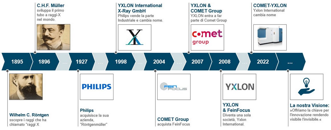
The listed sources provide a solid foundation for the presented information and are available for detailed verification of the claims made in the text.----------------------------------------------------------------------------------------------------------------------XRAYCONSULT, as the representative of Comet-Yxlon in Italy, is ready to provide assistance for all your radiographic needs.The Evolution of Industrial Inspection from the Beginning to Today by Comet-YxlonThe history of Comet-Yxlon in the field of industrial X-ray inspection began in 1895 when the German physicist Wilhelm Conrad Röntgen discovered this revolutionary technology.His discovery opened new possibilities in medical diagnosis and industrial inspection. However, it was in 1896 that the German entrepreneur Carl Heinrich Florenz Müller played a key role in developing the first X-ray tube, transforming Röntgen's discovery into a practical application.In 1927, Müller's factory was acquired by Philips. A pivotal moment in Comet-Yxlon's history was the collaboration with Philips in 1998. During that period, Philips decided to focus primarily on the medical sector, separating from the industrial branch.This decision led to the creation of Yxlon International GmbH, specializing in industrial X-ray inspection.The growth path didn't stop there. In 2007, Yxlon was acquired by the Comet Group, a global leader in the production of metal-ceramic X-ray tubes.This union resulted in the merger with Fein Focus, previously acquired by Comet in 2004, strengthening Yxlon's position in X-ray inspection and computerized tomography (CT), significantly contributing to quality and safety in various industrial sectors.In 2022, after 95 years of history, Yxlon International changed its name to Comet-Yxlon, maintaining its position as a pioneer and leader in industrial X-ray inspection.Until now, Comet-Yxlon has consistently focused on improving its tomography facilities. In the near future, there may even be opportunities to explore applications involving innovative nanotube technology.

Comet-Yxlon: Evolution of Industrial Inspection from the Beginning to TodayOUR FACILITIESIn the current industrial landscape, characterized by increasing global competition, there is a need to adopt innovative technologies to remain competitive.In this context, X-ray computed tomography stands out for its revolutionary potential, providing detailed information that allows exploration of defects and imperfections otherwise challenging to detect.This advanced analytical capability is opening new avenues for innovation and improvement in industrial design and production.Our range of Comet-Yxlon systems addresses these ever-evolving needs, offering cutting-edge solutions for quality control and advanced analysis – key elements for excelling in the current competitive environment.We look to the future with confidence, aware that technological innovation is crucial to tackling today's challenges and seizing new opportunities for growth and development."Impianto di Micro-Tomografia Comet-Yxlon"La Xrayconsult is always available to provide information about this technology. To view our facilities, click on the specified link: Tomografia Industriale